


Founded by veterinary Surgeon Dr. Kenneth Sinibaldi, ASOC was one of the first exclusively surgical practices in the region. It has grown over the years and can now be found at its current location in Shoreline — a modern,
well-equipped facility with eight exam rooms and five state-of-the-art surgical suites. It has since become co-owned by four of the five
board-certified surgeons, who boast over 75 years of combined
experience. They're accompanies by a professional support staff of
licensed veterinary technicians, assistants and administrators.
We are entirely focused on surgery, orthopedic medicine and rehabilitation, with unrivaled expertise. When you walk through our door, our overarching goal is to quickly establish an accurate diagnosis, perform only the necessary treatments, care for your pet through recovery, and restore them to optimal health. We’re committed to providing superb service to you, your pet, and your primary care veterinarian throughout the entire process. We are locally owned and operated, with our owners working in the practice full time. Our priority is to personally serve our patients and our community.
For more information about our rehabilitation services, visit SOUND Veterinary Rehabilitation Center.


A veterinary surgeon has undergone additional training after veterinary school in order to become a specialist. This training consists of a minimum of a 1-year internship followed by a 3-year residency program that meets guidelines established by the American College of Veterinary Surgeons (ACVS).
During the residency, there are specific training and caseload requirements that must be met. In addition to these requirements, applicants must perform research that is published in a scientific journal and then pass a rigorous examination. Specialists are called a “Diplomat of the American College of Veterinary Surgeons” or a “board-certified surgeon.”
ASOC is made up of a team of individuals who are effortlessly friendly, service-oriented, and highly trained. We’re a proud collective of five board-certified surgeons supported by devoted support staff.
Meet The Team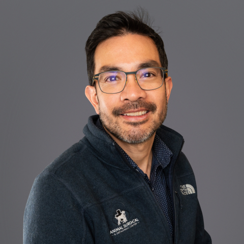
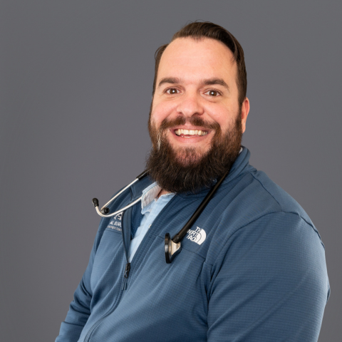
Alex Aguila, DVM, DACVS
Surgeon, Owner
Alex Aguila, DVM, DACVS
Diplomate ACVS
Surgeon, Owner
Dr. Alex Aguila completed his veterinary degree at the New York State College of Veterinary Medicine at Cornell University in 2000. Following veterinary school, he completed a year-long internship in small animal medicine and surgery in Springfield, Massachusetts, at Rowley Memorial Animal Hospital, and a year-long surgical internship in Pattersonville, New York, at the Capital District Veterinary Surgical Associates. Dr. Aguila then returned to Cornell, where he completed a three-year residency in small animal surgery. He earned board certification in small animal surgery in 2006.
Prior to joining Animal Surgical in 2008, Dr. Aguila gained substantial experience in a variety of surgical procedures while in private practice at Bay Area Veterinary Specialists in Houston. He has lectured extensively to the veterinary community, both in academic and private practice settings, and his published works include studies exploring the biomechanics of locking compression plates. His professional interest includes cranial cruciate ligament deficiency and minimally invasive surgery including fracture repair and locking plate technology. In his free time, Dr. Aguila likes to spend time with his family, getting outside as often as possible to go hiking, camping, skiing, running and expanding his newfound interest in cycling.
Allen Johnson, DVM, DACVS
Surgeon, Owner
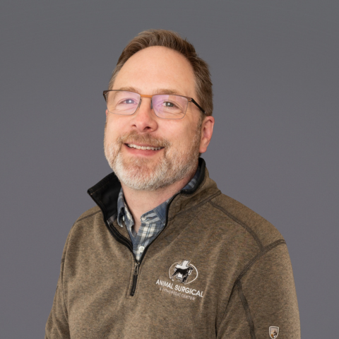
Allen Johnson, DVM, DACVS
Diplomate ACVS
Surgeon, Owner
Dr. Allen Johnson received his Doctor of Veterinary Medicine degree from Colorado State University in 1996, then completed a one-year rotating internship at Angell Memorial Animal Hospital in Boston, Massachusetts. He went on to complete a three-year surgical residency at Michigan State University, earning board certification in 2001.
Following his residency, Dr. Johnson remained at Michigan State University for two years as an assistant professor in small animal orthopedic surgery. While there, he was integral in helping develop MSU’s Canine Arthroscopy program. He moved to Seattle to join Animal Surgical in 2002.
Dr. Johnson’s research has been published in several veterinary journals and texts, and he has lectured at national and international veterinary surgical meetings. His professional interests include canine arthroscopy, patellar luxation, correction of angular limb deformities, and distraction osteogenesis.
Outside of work, he enjoys golfing, hiking, and exploring the Northwest with his wife and three sons. They share their home with their Golden Retriever, Annie, and a pointer mix named Frankie.
Jeff Mitchell, DVM, DACVS
Surgeon, Owner
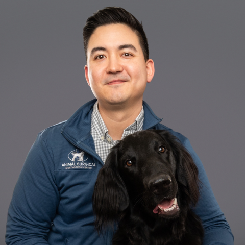
Jeff Mitchell, DVM, DACVS
Diplomate ACVS
Surgeon, Owner
Dr. Jeff Mitchell completed his doctorate degree in Veterinary Medicine from Washington State University. Shortly after this, he finished a small animal internship at the College of Veterinary Medicine Texas A&M, before completing a residency program at UC Davis where he gained his board certification. Dr. Mitchell’s professional interests include fracture management, joint disease and arthroscopy. He is also adept at a wide array of minimally invasive surgical techniques.
While not at work you can find him spending time with his wife Mariah, and their dog, Arya. They enjoy camping, bike rides, cooking and going on road trips which is why life in the Pacific Northwest suites them both to a tee!
Mike Thoesen, DVM, DACVS
Surgeon, Owner
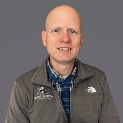
Mike Thoesen, DVM, DACVS
Diplomate ACVS
Surgeon, Owner
Dr. Mike Thoesen completed his Veterinary Medicine degree from Kansas State University in 2002. Following this, he completed a year-long internship at Purdue College of Veterinary Medicine. He then finished a residency program at Cornell University of Veterinary Medicine, gaining his board certification in 2007. Prior to joining Animal Surgical, Dr. Thoesen worked for 11 years in Omaha, Nebraska, at Midwest Veterinary Referral and Emergency Center. In addition to clinical practice, he taught fourth-year veterinary students, lectured extensively to the surrounding veterinary community, and published multiple cases and research papers.
His professional interests include regenerative bone healing and complex fracture management.
It seems that animal health is a family affair as his wife, Keli Anne, is also a veterinarian. Dr. Thoesen and his family started a new adventure in July of 2017 by moving to Seattle — his wife’s hometown. They have two daughters, a black Labrador named Emma, and two cats, Kenny and Chief.
In his free time, he enjoys spending time with his family, mountain biking, fishing, and camping.
Nick Rappa, DVM, DACVS
Surgeon
Nick Rappa, DVM, DACVS
Diplomate ACVS
Surgeon
Dr. Rappa earned his cum laude B.A. in Biochemistry, Cell, and Molecular Biology from Drake University before completing his DVM at Iowa State University, where he received two competitive scholarships. With extensive experience as a surgeon across top-tier veterinary specialty centers, he has also contributed to advancements in veterinary science through published research and studies on canine stifle repairs. Passionate about teaching and animal welfare, Dr. Rappa has mentored future professionals and volunteers with local humane societies to make a meaningful impact.
Desmond Tan, BVSc (Hons), DACVS
Surgeon
Desmond Tan, BVSc (Hons), DACVS
Surgeon - photo coming soon!
Surgeon
Dr. Tan earned his Bachelor of Veterinary Science with First Class Honors from the University of Melbourne. With extensive experience across internationally recognized veterinary centers, he has contributed to advancements in the field through research on orthopedic techniques, minimally invasive approaches, and innovative 3D-printed solutions. Outside of his professional work, he enjoys scuba diving, exploring mountain landscapes, and spending time with his two dogs—Alfred and Nigel—and his two cats, Casper and Edward.
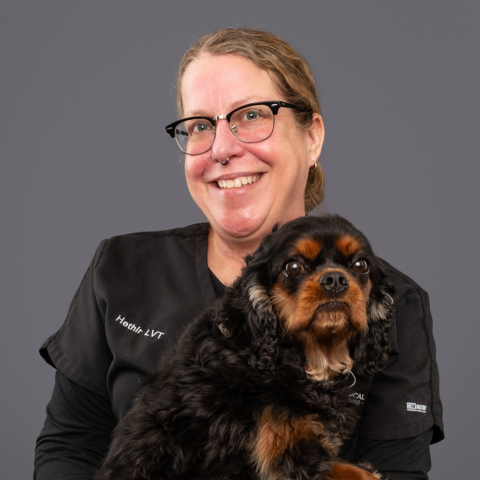
Hethir
Licensed Veterinary Technician
Hethir
Licensed Veterinary Technician
Hethir grew up in the Central Valley of California and attended Humbolt State University, majoring in literature and philosophy. She had a few stints at various jobs, including a barista and a projectionist at a small movie theatre, but it was working at a spay and neuter clinic during a gap year that set the course of her life. She knew that veterinary medicine was where her heart was, so she moved up to Seattle for the rain and the trees in 1998 and started working at The Animal Eye Clinic where she remained until April of 2017. Although she enjoyed her long, rewarding career at AEC, she was looking to challenge herself and since she always had an interest in surgery, she jumped at the chance to work for Animal Surgical when a position became available. And we’re very glad she did! She also enjoys cooking tasty vegan dishes, camping, traveling, hiking and taking long walks with her doggie.
.png)
Erika
Licensed Veterinary Technician
Erika
Licensed Veterinary Technician
Born in Seoul, Korea, and raised amidst the verdant landscapes of Puyallup, Washington, Erika Kim's journey into veterinary medicine was inevitable. With a heart drawn to those with four legs and a tail, she found her calling in caring for our furry companions. At home, Erika shares her space with a lively menagerie: 5 chickens, 2 dogs—Denna and Nandor—and a cat, each adding their own unique touch to her life. Outside the clinic, you'll often find her amidst nature, foraging for mushrooms alongside her faithful canine companions.
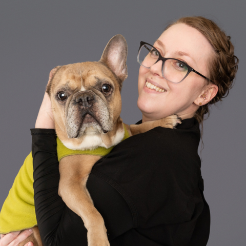
Gretchen
Licensed Veterinary Technician
Gretchen
Licensed Veterinary Technician
Gretchen started in the veterinary field in 2006 in the shelter system. She has a special interest in critical care medicine, and anything related to brachycephalic breeds. In her spare time she is usually painting, or posting too many Instagram pictures of her Frenchie, Gingko.
Kianna
Licensed Veterinary Technician
Kianna
Licensed Veterinary Technician
Kianna came to ASOC with a unique background in nurse training and emergency medicine that has shaped her compassionate, patient-centered approach to veterinary medicine. She is dedicated to providing high-quality care while supporting both pets and their families, bringing strong clinical skills and a calm, caring presence to every patient she works with.
Sari
Licensed Veterinary Technician
Sari
Licensed Veterinary Technician
Sari started her first career as a vocalist, traveling throughout the US, Canada and many of the Caribbean Islands in a variety of venues. But her love of animals drew her to pursue veterinary medicine. She joined Animal Surgical in 2000 after receiving her AA from Harcum College and interning at the University of Pennsylvania. She particularly enjoys her role as a resident gardener. She is also in charge of instructing the many interns and externs that rotate through our clinic.
Sari shares her home with Ramona, a terrier mix, and Winnie, a mix of all kinds! She enjoys gardening, bicycling, paddle boarding, yoga, singing in the shower and impersonating her coworkers!
Allie
Surgical Coordinator & Licensed Veterinary Technician
Allie
Surgical Coordinator & Licensed Veterinary Technician
Allie is a Licensed Veterinary Technician with over 10 years of experience in veterinary medicine. Her professional interests include anesthesia and pain management, where she is passionate about ensuring patients receive safe, individualized care and exceptional comfort throughout their procedures. Outside of work, Allie enjoys creating stained glass masterpieces and exploring the great outdoors.
Leah
Licensed Veterinary Technician
Leah
Licensed Veterinary Technician
Leah was born in Spokane, WA, endowed with an innate love of animals. Her mother explained that ever since she could talk, she told everyone she met that she was going to be a vegetarian — meaning, veterinarian, of course, although she dabbled with the former as well.
A month after graduating high school she started the veterinary technician program at PIMA and became licensed in 2011. She worked in general practice for four years and volunteered at the Humane Society. During that time, she discovered her passion for surgery and anesthesia and felt her place was in the O.R. So when a position became available at our clinic, she jumped at the chance to become part of our team.
She says she loves what she does and has no doubt that she’s found her calling. Her love of specialty medicine is inspiring her to consider taking it a step further and become certified as a Veterinary Technician Anesthetist.
She shares her home with a 2-year-old rescued cattle dog mix named Jazz (a.k.a. Mr. Jazzy Fizzle), quite the diva of a Netherland Dwarf bunny rabbit named Ninja, and newlywed husband, Justin. She enjoys camping, spending time with family, lazy days, food and traveling. Ever the animal lover, she spent time volunteering for an animal rescue while in the Dominican Republic on her honeymoon!
_600px.jpg)
Jenny
Licensed Veterinary Technician
Jenny
Licensed Veterinary Technician
Jenny joined Animal Surgical in 2000 after receiving her AA from Bel-Rea Institute of Animal Technology in Denver and interning at Alameda East Veterinary Hospital. Her particular interests are radiology and critical care of the surgical patient. She is our in-house Pitbull expert (the dog breed, not the singer) and advocate, and she shares her home with Salvador, her handsome pitty rescue.
.png)
Courtney
Licensed Veterinary Technician
Courtney
Licensed Veterinary Technician
Courtney is no ordinary veterinary professional – she's a licensed veterinary technician with 14 years of dedicated service in the field. Her journey has been a unique blend of administrative excellence and hands-on compassion, making her a valuable asset to the world of animal care.
After dedicating 7 years to management roles in the veterinary industry, Courtney felt a calling to return to her roots and rekindle her love for direct patient care. This profound passion led her to pursue her veterinary technician license, allowing her to once again work hands-on with the animals she adores.
Deyanira
Assistant Lead
Deyanira
Assistant Lead
Deyanira is our Assistant Lead and has more than 12 years of experience in veterinary medicine. Originally from Ecuador, she is passionate about delivering exceptional patient care while supporting and mentoring her team. Outside of work, Deyanira enjoys spending time with her two rescue dogs, who keep her active and always bring a smile to her day.
.jpg)
Crystal
Veterinary Assistant
Crystal
Veterinary Assistant
Crystal always held an interest in science and animals since childhood. It was this that drove her to get a Bachelor of Science in Zoology from Washington State University in 2017. She went on to volunteer with raptors for three years and started working at various general practice clinics as a kennel assistant and then a veterinary assistant. Crystal started working for Animal Surgical in the Fall of 2020 as our overnight assistant and has never looked back. She loves caring for our post-op patients and ensuring their recovery is smooth and comfortable.
Outside of working Crystal enjoys spending time with her family dogs, Apollo and Katy, and has special interests in the plant world, and birding in the Springtime.
Jerrica
Veterinary Assistant
Jerrica
Veterinary Assistant
Jerrica was born and raised here in Washington. Animals have always been a big part of her life. Prior to working with us, Jerrica worked at a local general practice clinic. Looking to expand her knowledge and experience she made the decision to move to a more specialized field, which is when she hopped on the opportunity to work for Animal Surgical.
In her free time, she and her husband enjoy spending time with their four dogs, watching movies, camping, and traveling. Her favorite spot so far has been Disneyland!
Elena
Veterinary Assistant
Elena
Veterinary Assistant
Elena earned her bachelor's degree in Environmental Science and Terrestrial Resource Management. She primarily works as a doctor's assistant, where she enjoys supporting patients and collaborating closely with the veterinary team to provide exceptional care. Her previous experience as both a veterinary assistant and front desk team member gives her a well-rounded understanding of veterinary medicine allowing her to provide excellent care to our patients.
Shelby
Veterinary Assistant
Shelby
Veterinary Assistant
Patricia
Veterinary Assistant
Patricia
Veterinary Assistant
After a life-long admiration of animals, Tricia attended Sno-Isle Skill Center to become a veterinary assistant during her senior year of high school in 2013. A year after graduating, she got a job at a general practice and worked there for seven years, excelling as a cross-trained receptionist/veterinary assistant. Looking for a change of pace, she started at ASOC in 2021 and looks at the transition as an incredible decision. In her free time, she helps make short films (mostly horror or comedy) with her boyfriend who is an aspiring director. She also enjoys defeating her boyfriend in Mario Kart. Her cat, Coco Marley, is practically her daughter and is spoiled as such. Nothing brings her more joy than taking care of animals and she found the perfect fit here at ASOC.
Samantha
Veterinary Assistant
Samantha
Veterinary Assistant
Paul
Pack Room Manager
Paul
Pack Room Manager
Paul joined Animal Surgical in 1999. As the pack room manager, he is responsible for sterilization, maintenance and inventory control for all surgical instruments and equipment and making sure the surgeons have the necessary paraphernalia for the many procedures they perform. Paul’s quick wit and sense of humor lend a bit of comic relief in our somewhat serious environment! Paul enjoys traversing the trails of Discovery Park with his wife, Lisa, traveling to the United Kingdom and chilling on the couch with his kitty, Mina. Paul is also an avid reader of a wide range of literature, from Lord of the Rings to historical novels.

Paige
Veterinary Assistant
Paige
Veterinary Assistant
Bio to come!

Riley
Veterinary Assistant
Riley
Veterinary Assistant
Riley joined ASOC after working in the tech industry. Her father Dr. Patterson used to own ASOC before retiring, and so returning to her roots felt like a natural decision! She shares her home with Quincy a terrier x corgi mix. She one day hopes to pursue a career as a veterinary technician!

Dakota
Veterinary Assistant
Dakota
Veterinary Assistant
Dakota grew up in Montana and decided to move to Washington with his girlfriend Amanda, two dogs, Lilly and Sully as well as his pet snake Oreo. He has a big interest in film and has a weekly movie night and is happy to share his most recent ratings! He loves to talk and is very excited to join the ASOC assistant team!
Mayra
Veterinary Assistant
Mayra
Veterinary Assistant
Born and raised in Colombia, Mayra brings her passion for animals and dedication to helping others to every aspect of her work. As a graduate of Universidad La Salle - Bogota, Colombia, she is eager to expand her knowledge and skills in veterinary medicine here in the United States, with the goal of becoming a licensed veterinarian.
Mayra loves Italian food and is a proud cat mom to two feline companions. Her heart is filled by serving others, and she finds joy in making a difference in the lives of pets and their families.
Noelle
Practice Manager
Noelle
Practice Manager
Noelle has one of the longest tenures at Animal Surgical, joining the practice in 1997 as a referral coordinator. Her passion has always been creating lasting relationships with the clients and making sure that everything runs smoothly for the practice. Since becoming a Practice Manager in 2014, she became a Veterinary Management Institute Graduate, solidifying her position here at ASOC. She looks forward to continuing to help the practice to grow. Noelle and her husband spend their free time mountain biking and trying to keep up with their two Labradors, Wallice and Hitch.
Isabella (Issy)
Assistant Manager
Isabella (Issy)
Assistant Manager
Issy, short for Isabella, hails from Nottingham, England, where she inherited her love of animals, big and small. She has been working with animals since she was 17 years old. She became a certified Animal Care Assistant for the Royal Society for the Protection of Animals (RSPCA) and after completing her certification she moved to Thailand where she managed a small street animal clinic named PhaNgan Animal Care. It was there where she met her American-born husband, Chase, and the two of them took off to China where they both taught English to children in Beijing and Chongqing.
After a while, they both yearned to put down roots, so they moved to the Seattle area where her husband is originally from. She dove right back into the animal care field and worked at PAWS as Animal Care Lead before making her way to ASOC.
She may have settled down, but her adventurous spirit remains. She has climbed Mt. Rainier and is currently in training to become a marathon runner! She shares her love of corgis with The Queen and enjoys spending time with her spunky corgi, Arthur. She also enjoys cooking, trying exotic cuisines and traveling. She believes that if you dance for 10 minutes a day, you’ll increase your happiness by at least 50%. When you encounter Issy, be prepared to be charmed by her English accent and her infectious positivity!

Aubrey
Referral Coordinator
Aubrey
Referral Coordinator
Aubrey's roots trace back to a cozy corner of New Jersey, where they cultivated a passion for adventure. ASOC's welcoming environment and top-notch care, they felt like they had found their professional home. Outside of work, Aubrey thrives on the challenge of rock climbing, finding solace in the mountains and one day hopes to further their education in the veterinary field. They share their home with partner and two cats Ash and Teddy.
Kaitlyn
Front Desk Supervisor
Kaitlyn
Front Desk Supervisor
Kaitlyn was born and raised in Alabama before moving to Alaska as a teenager. This is where her love for the outdoors grew so, of course, Seattle was a top pick for her next destination. In her spare time, she enjoys photography, seeing new places and going on grand adventures. When she isn’t hitting the trails you can find her in the kitchen cooking up a storm or puzzling with her husband and two dogs, Vader and Kyder.
Prior to moving to Seattle she has worked in multiple veterinary clinics and has shown dogs since a very young age. Kaitlyn’s favorite animal is tied between Shibas and Otters, although to some it’s more of an obsession than an interest.
Animal Surgical and Orthopedic Center has successfully treated thousands of animals thanks to our team’s depth of knowledge and commitment to their patients. Each day we vow to stay loyal to our mission statement.


Please note that Animal Surgical & Orthopedic Center sees patients by referral only. You can give us a call during our regular business hours (Monday - Friday, 8am - 6pm) or request an appointment online. We look forward to seeing you!
Request an Appointment


ASOC Clinic Hours
Monday to Friday: 8 AM – 6 PM
Saturday and Sunday: Closed
Major Holidays: Closed
Copyright © 2022
Animal Surgical And Orthopedic Center.
All Rights Reserved.
Website made by Wink
